


Two redundant transcription factor binding sites in a single enhancer are essential for mammalian sex determination
Meshi Ridnik , Elisheva Abberbock, Veronica Alipov, Shelly Ziv Lhermann, Shoham Kaufman, Maor Lubman, Francis Poulat, Nitzan Gonen

Genetics, cell biology and development
In mammals, the chromosomal sex (XX or XY) is established at fertilization. However, the undifferentiated embryonic gonad, or genital crest, remains bipotential until the stage of gonadal determination, at which point it differentiates into either an ovary or a testis.
This event occurs at approximately eleven days of embryonic life in mice and at approximately five weeks of gestation in humans.
 The choice between the testicular and ovarian pathways is made in undifferentiated precursors of the genital crest, which differentiate into supporting cells: the testicular Sertoli cells or the ovarian pre-granulosa cells.
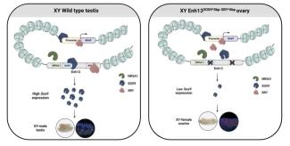
In XY gonads, the expression of SRY, which is carried by the Y chromosome, activates the SOX9 gene, which controls Sertoli cell differentiation.
 In XX gonads, the -KTS splice variant of WT1 associated with the WNT4/RSPO1 pathway leads to expression of the transcription factor FOXL2, which controls pre-granulosa cell differentiation.
Following their determination, the supporting cells orchestrate the development of the gonads, resulting in functional organs that produce sex hormones and gametes after puberty. Any disruption to these processes can result in disorders of sexual development, often associated with infertility, which can in turn lead to complete sex reversal.
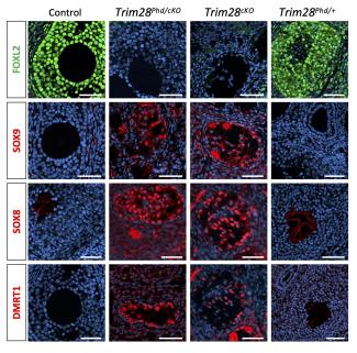
 Furthermore, supporting cells play a crucial role in maintaining the sexual identity of adult gonads. Our recent research has revealed that targeted deletion of the Trim28 gene in ovarian somatic cells induces their post-natal reprogramming into testicular cells. The process entails the trans-differentiation of granulosa cells into Sertoli cells, which ultimately leads to postnatal gonadal sex reversal.
In addition to studying testicular and ovarian determination processes, our group uses murine genetics to elucidate the mechanisms of differentiation and maintenance of the adult sexual phenotype. To this end, we use histological, transcriptomic, genomic and biochemical approaches to study our mouse models.
Meshi Ridnik , Elisheva Abberbock, Veronica Alipov, Shelly Ziv Lhermann, Shoham Kaufman, Maor Lubman, Francis Poulat, Nitzan Gonen
Isabelle Stevant Nitzan Gonen Francis Poulat
Pascal Philibert, Stéphanie Déjardin, Mélissa Girard, Quentin Durix, Anne-Alicia Gonzalez, Xavier Mialhe, Mathieu Tardat, Francis Poulat, Brigitte Boizet-Bonhoure
Moïra Rossitto , Stephanie Déjardin , Chris M Rands , Stephanie Le Gras , Roberta Migale , Mahmoud-Reza Rafiee , Yasmine Neirijnck , Alain Pruvost , Anvi Laetitia Nguyen , Guillaume Bossis , Florence Cammas , Lionel Le Gallic , Dagmar Wilhelm , Robin Lovell-Badge , Brigitte Boizet-Bonhoure , Serge Nef , Francis Poulat
B. Boizet-Bonhoure, S. Dejardin, M. Rossitto, F. Poulat and P. Philibert
Rossitto M, Ollivier M, Déjardin S, Pruvost A, Brun C, Marchive C, Nguyen AL, Ghettas A, Keime C, de Massy B, Poulat F, Philibert P, Boizet-Bonhoure B
Weger BD, Gobet C, Yeung J, Martin E, Jimenez S, Betrisey B, Foata F, Berger B, Balvay A, Foussier A, Charpagne A, Boizet-Bonhoure B, Chou CJ, Naef F, Gachon F
van Dijk J, Bompard G, Cau J, Kunishima S, Rabeharivelo G, Mateos-Langerak J, Cazevieille C, Cavelier P, Boizet-Bonhoure B, Delsert C, Morin N
Rossitto M, Marchive C, Pruvost A, Sellem E, Ghettas A, Badiou S, Sutra T, Poulat F, Philibert P, Boizet-Bonhoure B
Gonen N, Futtner CR, Wood S, Garcia-Moreno SA, Salamone IM, Samson SC, Sekido R, Poulat F, Maatouk DM, Lovell-Badge R
Penrad-Mobayed M, Perrin C, L'Hôte D, Contremoulins V, Lepesant JA, Boizet-Bonhoure B, Poulat F, Baudin X, Veitia RA
Rahmoun M, Lavery R, Laurent-Chaballier S, Bellora N, Philip GK, Rossitto M, Symon A, Pailhoux E, Cammas F, Chung J, Bagheri-Fam S, Murphy M, Bardwell V, Zarkower D, Boizet-Bonhoure B, Clair P, Harley VR, Poulat F.
Boizet-Bonhoure, B.
Rossitto M, Philibert P, Poulat F, Boizet-Bonhoure B.
Rossitto M, Ujjan S, Poulat F, Boizet-Bonhoure B
Trimarco A, Forese MG, Alfieri V, Lucente A, Brambilla P, Dina G, Pieragostino D, Sacchetta P, Urade Y, Boizet-Bonhoure B, Boneschi FM, Quattrini A, Taveggia C.
Rahmoun M, Perez J, Saunders PA, Boizet-Bonhoure B, Wilhelm D, Poulat F, Veyrunes F.
Moniot B, Ujjan S, Champagne J, Hirai H, Aritake K, Nagata K, Dubois E, Nidelet S, Nakamura M, Urade Y, Poulat F, Boizet-Bonhoure B
Philibert P, Boizet-Bonhoure B, Bashamboo A, Paris F, Aritake K, Urade Y, Leger J, Sultan C, Poulat F.
Lichten, M., de Massy, B.
Moniot B, Farhat A, Aritake K, Declosmenil F, Nef S, Eguchi N, Urade Y, Poulat F, Boizet-Bonhoure B.
Sim H, Argentaro A, Czech DP, Bagheri-Fam S, Sinclair AH, Koopman P, Boizet-Bonhoure B, Poulat F, Harley VR
Dumond, H., Al-Asaad, I., Chesnel, A., Chardard, D., Boizet-Bonhoure, B., Flament, S., Kuntz, S.
Farhat A, Philibert P, Sultan C, Poulat F, Boizet-Bonhoure B.
Abdel-Samad R, Zalzali H, Rammah C, Giraud J, Naudin C, Dupasquier S, Poulat F, Boizet-Bonhoure B, Lumbroso S, Mouzat K, Bonnans C, Pignodel C, Raynaud P, Fort P, Quittau-Prévostel C, Blache P
Lakhdar R, Denden S, Knani J, Leban N, Daimi H, Hassine M, Lefranc G, Ben Chibani J, Haj Khelil A.
+
IMGT® - the international ImMunoGeneTics information system®
Malki S, Boizet-Bonhoure B, Poulat F
Kalfa N, Méduri G, Philibert P, Patte C, Boizet-Bonhoure B, Thibaut E, Pienkowski C, Jaubert F, Misrahi M, Sultan C.
Malki S, Boizet-Bonhoure B, Poulat F
Kalfa, N., Veitia, RA., Benayoun, BA., Boizet-Bonhoure, B., Sultan, C.
Kalfa, N., Philibert, P., Sultan, C
Kalfa, N., Philibert, P., Sultan, C.
Moniot, B., Declosmenil, F, Barrionuevo, F., Scherer, G., Aritake, K., Malki, S., Marzi, M., Cohen-Solal, A., Georg, I., Klattig, J., Englert, C., Kim, Y., Capel, B., Eguchi, N., Urade, Y., Boizet-Bonhoure, B., Poulat, F.
Kalfa N, Philibert P, Patte C, Thibaud E, Pienkowski C, Ecochard A, Boizet-Bonhoure B, Fellous M, Sultan C.
Moniot, B., Boizet-Bonhoure, B., Poulat, F.
Malki, S., Declosmenil, F., Farhat, A., Moniot, B., Poulat, F., Boizet-Bonhoure, B.
Kalfa, N., M. Fellous, B. Boizet-Bonhoure, C. Patte, P. Duvillard, C. Pienkowski, F. Jaubert, A. Ecochard and C. Sultan.
El Jamil, A., Kanhoush, R., Magre, S., Boizet-Bonhoure, B., Penrad-Mobayed, M.
Manuylov, NL., Fujiwara, Y., Adameyko, II., Poulat, F., Tevosian, SG.
Malki, S., Bibeau, F., Notarnicola, C., Roques, S., Berta, P., Poulat, F., Boizet-Bonhoure, B.
Galmiche, L., Sarnacki, S., Verkarre, V., Boizet, B. Duvillie, B., Fabre, M., Jaubert, F
Köhler, B., Delezoide, AL., Boizet-Bonhoure, B., McPhaul, MJ., Sultan, C., Lumbroso, S
Kim, Y., Kobayashi, A., Sekido, R., DiNapoli, L., Brennan, J., Chaboissier, M.C., Poulat, F., Behringer, R.R., Lovell-Badge, R. and Capel, B.
Notarnicola C, Malki S, Berta P, Poulat F, Boizet-Bonhoure B.
Notarnicola C, Malki S, Berta P, Poulat F, Boizet-Bonhoure B.
Malki, S., Berta, P., Poulat, F., Boizet-Bonhoure, B.
Malki, S., Nef, S., Notarnicola, C., Thevenet, L., Gasca, S., Méjean, C., Berta, P., Poulat, F., Boizet-Bonhoure, B.
Nef, S., Schaad, O., Stallings, N.R., Cederroth, C.R., Pitetti, J.L., Schaer, G., Malki, S., Dubois-Dauphin, M., Boizet-Bonhoure, B., Descombes, P., Parker, K.L. and Vassalli, J.D.
Thevenet, L., Albrecht, K.H., Malki, S., Berta, P., Boizet-Bonhoure, B. and Poulat, F.
Thevenet, L., Mejean, C., Moniot, B., Bonneaud, N., Galeotti, N., Aldrian-Herrada, G., Poulat, F., Berta, P., Benkirane, M., Boizet-Bonhoure, B.
Moniot, B., Biau, S., Faure, S., Nielsen, C.M., Berta, P., Roberts, D.J. and de Santa Barbara, P.
Zhou, R., Bonneaud, N., Yuan, C., de Santa Barbara, P., Boizet, B., Schomber, T., Scherer, G., Roeder, G., Poulat, F., Berta, P.
Hardelin, J.P., Julliard, A.K., Moniot, B., Soussi-Yanicostas, N., Verney, C., Schwanzel, M., Ayer-Le-Lièvre, C. and Petit, C.
Defended by Moïra Rossitto on 09-12-2016